Reflection XVI: Glycolysis I

Hey guys,

Today we begin glycolysis! YAYYY!!!

For this reflection,  I have posted biochemjm’s (hokage’s) first video above.


Summary

Glycolysis

In summary, a single glucose molecule in glycolysis produces a total of 2 molecules of pyruvic acid, a net gain of 2 molecules of ATP, 2 molecules of NADH and 2 molecules of water.

Although 2 ATP molecules are used in steps 1-3, 2 ATP molecules are generated in step 7 and 2 more in step 10. This gives a total of 4 ATP molecules produced. If you subtract the 2 ATP molecules used in steps 1-3 from the 4 generated at the end of step 10, you end up with a net total of 2 ATP molecules produced.

I also found this animation of glycolysis to be particularly helpful 😀

http://highered.mcgraw-hill.com/sites/0072507470/student_view0/chapter25/animation__how_glycolysis_works.html

Hope you guys learnt stuff 🙂

Reflection XV: INHIBITION DUN DUN DAHHHHHHH X__X

INHIBITION!

INHIBITION EVERYWHERE!!!

Okay maybe I’m over reacting, but think about it. Imagine if you were an enzyme, minding your own business and BAM! some random molecule comes and changes you completely leaving you unable to function.

I mean it isn’t very nice especially if you’re a particularly helpful enzyme. Of course not all inhibition leads to this fate.

I guess it really is a perspective thing though. Because if you think about it,  the inhibition of enzymes may not seem nice from an enzyme perspective but what if that enzyme is catalyzing an unwanted or unnecessary reaction? The body actually utilizes a certain type of inhibition as a from of negative feedback to prevent the production of excess products and a waste of cellular reserves.

We ALL know where this is going, so I guess we should get right into it.

Enzyme inhibitors are molecules that interfere with catalysis by slowing down of halting enzymatic reactions. Inhibitors can affect enzymes either reversibly or irreversibly.

Inhibition

REVERSIBLE INHIBITION

Reversible enzyme inhibition does not permanently alter the enzyme and usually occurs when reversible inhibitors bind non-contently with the enzyme, resulting in four different types of reversible inhibition.

Competitive inhibition occurs when a competitive inhibitor is present. This inhibitor molecule, which is structurally similar to the substrate, competes for the active site of the enzyme by occupying it, forming an enzyme inhibitor complex and preventing the substrate from binding to the enzyme active site. This can be further analyzed via the following graph:

Lineweaver Burke Plot of Reversible Inhibition: Competitive Inhibition (Bliq. 2013)

In the presence of a competitive inhibitor Vmax of the reaction remains constant, however, Km of the reaction increases as it takes a higher concentration of the substrate to reach half the Vmax. The effects if competitive inhibition can be lowered by increasing the concentration of substrate.

Uncompetitive inhibition, another type of reversible inhibition, occurs when  the inhibitor, which does not resemble the substrate, binds to a site distinct from the active site and binds only to the enzyme-substrate complex. This causes the Vmax to decrease as a result of removing the activated complex and thus decreases Km.This can be observed in the graph blow.

Lineweaver Burke Plot of Reversible Inhibition: Uncompetitive Inhibition (Bliq. 2013)


Mixed inhibition is the third type on reversible inhibition and arises from the presence of an inhibitor, that does not resemble the substrate, which binds to a site distinct from the active site on either the enzyme-substrate complex or the enzyme itself. Both Vmax and Km are affected. Vmax is lowered while Km increases. This can be seen in the graph:

Lineweaver Burke Plot of Reversible Inhibition: Mixed Inhibition (Bliq. 2013)

 The last type of reversible inhibition is noncompetitive inhibition which occurs when the binding of the inhibitor, which does not structurally resemble the substrate, to the enzyme reduces enzyme activity but does not affect the enzyme-substrate binding; therefore, as a result the extent of inhibition is dependent on the concentration of the inhibitor, i.e. as the concentration of the inhibitor increases the slower the rate of reaction. Hence in the presence of these inhibitors Vmax decreases and Km remains the same. This can be seen in the graph below.

Lineweaver Burke Plot of Reversible Inhibition: Noncompetitive Inhibition (Bliq. 2013)


IRREVERSIBLE INHIBITION

  Irreversible enzyme inhibition occurs when the inhibitor interacts with the enzyme and changes it chemically at the active site. This usually occurs by bonding covalently with the enzyme, destroying a functional group that is essential to ezymatic activity or by forming a particularly stable non covalent enzyme-inhibitor complex. 

Irreversible Inhibition

Welp thats all for now folks ^_^

Reflection XIV: Isozymes ^_^

Hey Biochemians,

Today we’re going to look at isozymes. Although not gone into detail in the course, when I was checking the course outline, I happened to notice that it was one of the terms we needed to know.

Now before we walk into the exam not knowing,

or panicking,

I shall now briefly explain the fun and importance that is isozymes. ^_^

Isozymes (also known as isoenzymes) are homologous enzymes that catalyze the same reaction but differ in structure. The differences in the isozymes allow them to regulate the same reaction at different places in the specie. These enzymes differ in amino acid sequences and display different kinetic parameters as well as regulatory properties.

Isozymes are encoded by different genes and expressed in a distinct organelle or at a distinct stage of development. The purpose of these isomers is to allow fine adjustment of metabolism to meet the need of different development stages and help the different tissues and organs function properly depending on their physiology and in what kind of environment which they function. Hence these enzymes appear in specific regions of the body; differing in specifics organelles or tissues.

And that’s the generalization of isozymes.

Happy studing

Reflection XIII: Inorganic Catalysts vs. Biological Catalysts

Hey guys 😀

Today I would like to reflect on the differences between inorganic catalysis and biological catalysts (enzymes).

Enzymes and inorganic catalysts both affect the rate of a reaction. The difference between the two is that, enzymes are largely organic in nature and are bio-catalysts.  Hence even though all known enzymes are catalysts, all catalysts are not enzymes. 

Here’s  a table which quite nicely sums up the basic comparisons.

Some things that I would like to add though to this are:

  • Enzymes are much more efficient than inorganic catalysts as inorganic catalysts usually require high temperatures and/or high pressures for the reactions to be catalyzed, whereas in our bodies hundreds and thousands of catalysts exist and perform at average body temperature (35-37 degrees Celsius). 
  • The catalytic power of enzymes is quite higher than that of inorganic catalysts.
  • Inorganic catalysts can be poisoned, easily whereas organic catalysts are not easily poisoned.
  • Enzymes are regulated, inorganic catalysts are not.

Yup that’s all for now folks.

Reflection XII: Rates! Rates! Rates

 Hey guys!

Well today, I’m going through the differing factors which affect rate of an enzyme catalyzed reaction. Paying specific attention to my three favorite factors.

The rate of ezymatic activity can be affected by many factors. These include enzyme concentration, substrate concentration, temperature, pH and presence of inhibitors.

The effect of substrate concentration on enzyme catalyzed reactions can be seen in the graph below.

Plot of Effect of Substrate Concentration on Initial Velocity of an Enzyme Catalyzed Reaction

    As graphically represented above, at the beginning of the graph, the rate of the reaction is proportional to substrate concentration; i.e. as the concentration of substrate increases  so does the rate. However, at higher substrate concentrations, velocity increases by smaller and smaller amounts; this leads to a plateau like region of the curve close to the maximum velocity (Vmax). This occurs due to the concentration of substrate being so high that all the active sites of enzymes are occupied and the enzymes are said to be saturated, hence the curve levels off.

   The effect of temperature on enzyme catalyzed reactions can be seen in the graph below.

Plot of Temperature on Initial Velocity of an Enzyme Catalyzed Reaction

As graphically represented above, at the beginning of the graph, the rate of the reaction is proportional to temperature increase; i.e. as the temperature increases  so does the rate due to increase in kinetic energy hence increasing the number of enzyme-substrate collisions. This goes on until an optimum temperature is observed. This is graphically represented by the highest point of the graph. However, at higher temperatures, beyond optimum temperature, velocity drops; this leads to the formation of a symmetrical curve. This occurs due to the denaturation of the enzyme at temperatures higher than optimum, where the inter-molecular forces which fold the protein are overcome and tertiary and quaternary structure’s of the enzyme are lost, hence the enzyme becomes inactive, slowing the rate of reaction.

Thirdly, pH is another limiting factor and can be represented graphically as:

Graphical Representation of Effect of pH on Enzyme Activity

As observed in the graph the rate of enzymes have an optimum pH or pH range at which their activity is maximum; This is referred to as the optimum pH and is specific for enzymes, based on where they are found. For instance the optimum pH range of enzymes found in the stomach is 2-3, while the optimum pH range of enzymes found in the intestine is 7-8.  At higher or lower pH values of the optimum, enzymatic activity decreases. This is because amino acid side chains in the active site with critical functions that depend on their maintaining a certain state of ionization may act as weak acids and bases hence can no longer act as the active site, hence can no longer catalyze reactions.

Enzymes are pH specific due to the

Reflection XI: Enzyme Assistance

Hey biochemians 🙂

Today I’m going to reflect on the chemical substances or ions that aid enzymes. We need to remember these basic and important definitions in order to move further ahead next semester. So when sir says “This enzyme has 5 cofactors….” we won’t be all confused like

First I guess to get a clear visual of these  we should define enzymes.

Enzymes are biological catalyst that speeds up chemical reactions by providing an alternative pathway with a lower activation energy.

Cofactors are non protein chemical compound that is required for the enzyme’s biological activity.

-The prosthetic group is the coenzyme or metal ion that is very tightly or covalently bonded to enzyme protein. 


-A holoenzyme is a complete catalalytically active enzyme together with its bound coenzyme and/or metal ions

– The apoenzyme is the protein part of the holoenzyme.

Apoenzyme + Prosthetic Group = Holoenzyme

Hope this helps you out guys! Rewriting it and seeing it in pictures helped me.

Happy studying 🙂

Reflection X: International Enzymes

Hey guys,

So today when studying I decided to come up with a mnemonic device to help us remember the six classes of enzymes.

The six classes are as follows:

  1. Oxidoreductases – Involved in the transfer of electrons
  2. Transferases – Involved in group transfer reactions
  3. Hydrolases – Involved in hydrolysis reactions
  4. Lyases – Involved in the addition of groups to double bonds or formation of double bonds by removal of groups
  5. Isomerases – Involved in the transfer of groups within molecules to yield isomeric forms
  6. Ligases – Involved in the formation of C-C, C-S, C-O, and C-N bonds by condensation reactions coupled to cleavage of ATP or similar co-factor.

Ok. Time for some imagination

IMAGINATION TIME

Old Trained Horses Lead Interesting Lives.

Guys I think my imagination may be broken :/

Though its not that hard to remember.

Yea, I think I’ll keep it. 🙂

Well, mission complete for today. You guys can try to remember this or come up with your own. Let me know how it goes.

Happy studying 😀

Reflection IX: Turnover? Turnunder?

HI GUYS 😀

So while I was studying and reading and stuff, making my way through the syllabus, I came across this new term I thought I would get a little more into; (especially since mentioned in one of the lectures that this is a favorite mcq) 😀

We all need to be prepared right? ^_^

The TURNOVER NUMBER a.k.a. kcat

The number of molecules of substrate converted to product per enzyme molecule per second  is referred to as the turnover number (also termed kcat) and can be calculated as follows: kcat = Vmax/[E]T.

Reflection VIII: Enzymes vs. Ribosymes

Hey guys!! 😀

 Most enzymes are proteins, but there are a small group of catalytic RNA molecules which are referred to as ribozymes.

A ribonucleic acid enzyme, or ribozyme is an RNA molecule that is capable of catalyzing specific biochemical reactions. The discovery of ribozymes contributed greatly to the RNA world hypothesis, which suggests that RNA may have been important in the evolution of prebiotic self-replicating systems.

Ribozymes

I came across this topic in class some time ago and again when I was studying. From what I knew, all biological catalysts were enzymes and these were proteins; So I decided to make this post to clarify and make a mental note and also to help out you guys, my fellow biochemians.

Hope its clear 🙂

Til next time.. Happy studying

Reflection VII: AMINO ACIDS & PROTEINS P2!!!

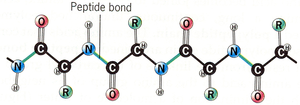
Amino Acids form undergo a condensation reaction to form a dipeptide.

Formation of Peptide Bond

This dipeptide can remain or link with more amino acids to form a polypeptide.

Polypeptide Chain

-The unique sequence of amino acids in the  polypeptide chain give rise to unique proteins that code for many things such as hair, skin, nails and enzymes.

Structure-Function Relationship

-Polypeptides must be folded to be functional proteins.

-Protein structure dictates protein function.

-Proteins have four levels of structure.

  • Primary Structure
  • Secondary Structure
  • Tertiary Structure
  • Quaternary Structure

Primary Structure.

-This the sequence of amino acid units, and comprises the protein’s overall biomolecular structure.

Primary Protein Structure

Secondary Structure 

This is the specific geometric shape caused by intramolecular and intermolecular hydrogen bonding of amide groups. Consists of two types alpha helix and beta pleated sheet.

Secondary Structure of Proteins

Tertiary Structure

This is the final specific geometric shape that a protein assumes. This is determined by a variety of bonding interactions between the “side chains” on the amino acids. These bonding interactions may be stronger than the hydrogen bonds between amide groups holding the helical structure. As a result, bonding interactions between “side chains” may cause a number of folds, bends, and loops in the protein chain. Different fragments of the same chain may become bonded together.

There are four types of bonding interactions:

  1. Disulfide bonds are formed by oxidation of the sulfhydryl groups on cysteine. Different protein chains or loops within a single chain are held together by the strong covalent disulfide bonds.
  2. Hydrogen bonding between “side chains” occurs in a variety of circumstances. The most usual cases are between two alcohols, an alcohol and an acid, two acids, or an alcohol and an amine or amide.
  3. Salt bridges/ Ionic bonds result from the neutralization of an acid and amine on side chains. The final interaction is ionic between the positive ammonium group and the negative acid group. Any combination of the various acidic or amine amino acid side chains will have this effect.
  4.  Non-Polar Hydrophobic Interactions: are believed to contribute significantly to the stabilizing of the tertiary structures in proteins. This interaction is really just an application of the solubility rule that “likes dissolve likes”. The non-polar groups mutually repel water and other polar groups and results in a net attraction of the non-polar groups for each other.

The four types of bonding that takes place within a protein’s tertiary structure that give it it’s geometrical shape.

Quaternary Structure

This involves the clustering of several individual peptide or protein chains into a final specific shape. A variety of bonding interactions including hydrogen bonding, salt bridges, and disulfide bonds hold the various chains into a particular geometry.

Quaternary Structure of Haemoglobin

There are two major categories of proteins with Quaternary structure:.

  1. Fibrous Proteins: the result of the interaction of many individual protein chains. Some are composed of hydrogen bonding on amide groups on different chains is the basis of beta-pleated sheet as seen in silk proteins.While other fibrous proteins such as keratins  are composed of coiled alpha helical protein chains with other various coils analogous to those found in a rope.  Fibrous proteins are insoluble, and play a structural or supportive role in the body, and are also involved in movement (as in muscle and ciliary proteins).
  2. Globular Proteins:  globular proteins fold back on themselves to produce compact, nearly spherical shapes. Most globular proteins are water soluble and hence are relatively mobile within a cell. Some examples are enzymes, antibodies, hormones, toxins, and substances such as hemoglobin.

Fibrous and Globular Proteins

Well that’s all I can remember; time to re-watch the lectures 😛

Good luck my fellow Biochemians